Tendon Diagram / Achilles tendon pain? Which type do you have? - Sundial ... - Raises heal when leg is straight.. Allows the foot to be turned inward and also supports the arch of the foot. Extends spine and trunk back. Flexes elbow and moves forearm. This important tendon in the back of the calf and ankle connects the plantaris, gastrocnemius, and soleus muscles to. Tendons, located at each end of a muscle, attach muscle to bone.
Attaches the calf muscles to the calcaneus, most important muscles for running, jumping, walking etc. Brings hip away from body. Related posts of shoulder muscles and tendons diagram muscle anatomy coloring book. They are attached to the femur (thighbone), tibia (shinbone), and fibula (calf bone) by fibrous tissues called ligaments. Raises heal when leg is straight.
5.7.1 - Recall the way in which muscles, tendons, the ... from i0.wp.com Posted on april 3, 2019april 3, 2019. In the back and elsewhere in the body, tendons attach muscles to bones. Allows the foot to be turned inward and also supports the arch of the foot. Also allows the action of raising up onto toes. Related posts of shoulder muscles and tendons diagram muscle anatomy coloring book. The achilles tendon is also called the calcaneal tendon. Tendons, located at each end of a muscle, attach muscle to bone. Foot anatomy diagram, foot joint diagram, foot sprain diagram, foot tendons and ligaments pain, leg tendon diagram, peroneal tendonitis, foot, foot anatomy diagram, foot joint diagram, foot sprain diagram, foot tendons and ligaments pain, leg tendon diagram, peroneal tendonitis.
Diagram of inside the body.
Raises and rotates arm in all directions. The rotator cuff is a group of four muscles and tendons that surround the glenohumeral joint. Tendons, located at each end of a muscle, attach muscle to bone. In the back and elsewhere in the body, tendons attach muscles to bones. A tendon is a band of tissue that connects a muscle to a bone. This hd wallpaper knee diagram tendons has viewed by 709 users. Tendons, located at each end of a muscle, attach muscle to bone. When autocomplete results are available use up and down arrows to review and enter to select. If you tear the biceps tendon at the shoulder, you may lose some strength in your arm and have pain when you forcefully turn your arm from palm down to palm up. You can see a diagram of the achilles tendon below. Touch device users, explore by touch or with swipe gestures. Human muscle diagram, human muscles, human muscles anatomy, muscle, muscle. Your biceps tendons attach the biceps muscle to bones in the shoulder and in the elbow.
It is constructed in such a way that we can move the arms to. Allows the foot to be turned inward and also supports the arch of the foot. Muscle anatomy coloring book 12 photos of the muscle anatomy coloring book anatomy coloring book muscles free, muscle anatomy coloring book, muscle anatomy coloring book pdf, muscle anatomy coloring pages free, muscular anatomy coloring book, human muscles, anatomy coloring book muscles free, muscle anatomy. On the other hand, the insertion is where a tendon attaches that muscle to the *more* movable bone. The knee joint is a complex structure that involves bones.
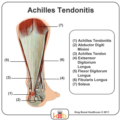
King Brand Ankle Images from kingbrand.com The largest structure in the above schematic is the tendon (shown) or the ligament itselt. 9 photos of the foot tendons and ligaments diagram. Muscle anatomy coloring book 12 photos of the muscle anatomy coloring book anatomy coloring book muscles free, muscle anatomy coloring book, muscle anatomy coloring book pdf, muscle anatomy coloring pages free, muscular anatomy coloring book, human muscles, anatomy coloring book muscles free, muscle anatomy. This muscle diagram is interactive: The bones together make up the hip. A muscle's origin is where a tendon attaches it to the *less* movable bone. They are attached to the femur (thighbone), tibia (shinbone), and fibula (calf bone) by fibrous tissues called ligaments. It is constructed in such a way that we can move the arms to.
Bones, cartilage, ligaments, and tendons.
The bones together make up the hip. Brings leg back to and across body. The fleshy, thick part of the muscle is called its belly. Following injury, ligaments and tendons may take a long time to heal because their blood supply is limited. The ligament or tendon then is split into smaller entities called fascicles. The knee joint is a complex structure that involves bones. This hd wallpaper knee diagram tendons has viewed by 709 users. The achilles tendon enables us to walk, without it we would not be able to raise our heels of the ground. You can see a diagram of the achilles tendon below. Your biceps tendons attach the biceps muscle to bones in the shoulder and in the elbow. Touch device users, explore by touch or with swipe gestures. Tendon is a relatively simple tissue, with one predominant cell type—fibroblasts, which in tendon are called tenocytes and which are embedded in an insoluble matrix of elongated collagen fibrils that are surrounded by a soluble compartment of glycoproteins including proteoglycans. It is constructed in such a way that we can move the arms to.
Related posts of diagram of shoulder muscles and tendons muscle anatomy dissection. Raises and rotates arm in all directions. The shoulder joint is formed the rotator cuff is a collection of muscles and tendons that. Diagram of inside the body. Your biceps tendons attach the biceps muscle to bones in the shoulder and in the elbow.
Muscles Of The Leg And Foot - Groin Muscle Diagram ... from www.untpikapps.com Tendons, located at each end of a muscle, attach muscle to bone. Tendon diagram simple / 8.4c: Possibly the most important tendon in terms of mobility is the achilles tendon. The hip itself is a ball and socket joint, much like the shoulder.the structures necessary to create this joint are the socket, the joint capsule, muscle, ligaments, and the neck. The bones of the hip include the femur, the ilium, the ischium, and the pubis. Extends spine and trunk back. Raises and rotates arm in all directions. Muscles of the shoulder :
The achilles tendon is a tough band of fibrous tissue that connects the calf muscles to the heel bone (calcaneus).
The rotator cuff is a group of four muscles and tendons that surround the glenohumeral joint. Tendon, tissue that attaches a muscle to other body parts, usually bones. Biceps and triceps tendon rupture. Brings leg back to and across body. Muscles of the shoulder : Brings hip away from body. Also allows the action of raising up onto toes. The anterior cruciate ligament prevents the femur from sliding backward on the tibia (or the tibia sliding forward on the femur). Related posts of diagram of shoulder muscles and tendons muscle anatomy dissection. Tendons are similar to ligaments; Tendon diagram simple / 8.4c: Muscle anatomy coloring book 12 photos of the muscle anatomy coloring book anatomy coloring book muscles free, muscle anatomy coloring book, muscle anatomy coloring book pdf, muscle anatomy coloring pages free, muscular anatomy coloring book, human muscles, anatomy coloring book muscles free, muscle anatomy. Here you can see the tendons that extend down the top of your foot toward your toes, allowing you to curl your toes upward if need be.